
In short
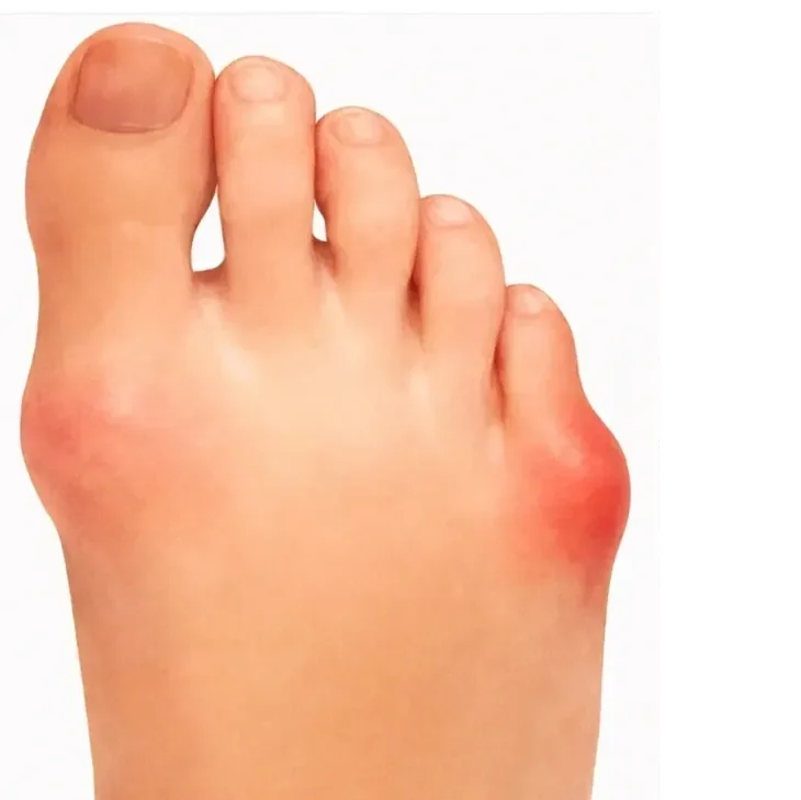
Bunionette surgery treats a painful prominence on the outside of the foot at the base of the little toe. Depending on the deformity, the bump is shaved (lateral eminence resection) or the 5th metatarsal is cut and repositioned (osteotomy), sometimes through minimally invasive surgery and sometimes fixed with small screws. You walk in a post-operative shoe, an X-ray at 6 to 12 weeks checks healing, and most people are back in normal footwear by then.
- Anaesthetic
- Local, regional or general anaesthetic
- Hospital stay
- Usually a day case
- Walking afterwards
- Protected weight-bearing in a post-operative shoe
- Full recovery
- 3 to 6 months
Why is surgery sometimes needed?
A bunionette, also known as a tailor’s bunion, is a prominence on the outside of the foot at the base of the little toe. This can cause pain, swelling and rubbing in shoes, especially narrower or firmer footwear. When symptoms persist despite conservative treatment, surgery may be recommended to realign the bone and reduce pressure.
What does the operation involve?
The procedure is usually performed as a day case under local, regional or general anaesthetic. A small incision is made over the bunionette, or it is done through minimally invasive surgery. Depending on the deformity, surgery may involve:
- Shaving the prominence (lateral eminence resection)
- Osteotomy (cutting and repositioning the bone)
- Fixation with small screws, if required
The soft tissues are balanced and the wound is closed with sutures. Dressings are applied and you may be given a post-operative shoe. The goal is to reduce the prominence and correct the alignment of the 5th metatarsal.
Bunionette surgery is an effective treatment for a painful prominence on the outer side of the foot that does not improve with conservative care. By correcting the underlying bone alignment and reducing the bony prominence, the procedure improves comfort, footwear tolerance and daily function. Most patients experience meaningful and lasting improvement.
Recovery: what to expect
0 to 2 weeks
- The foot is bandaged and protected in a post-operative shoe
- Protected weight-bearing is typically allowed unless instructed otherwise
- Elevation helps reduce swelling
- Stitches are removed at 10 to 14 days
2 to 6 weeks
- Swelling gradually settles
- Transition to comfortable footwear begins as swelling allows
- Walking distances may increase gradually
6 to 12 weeks
- An X-ray will be performed to check on healing progress
- Most patients return to normal footwear with good comfort
- Low-impact activities such as cycling and swimming may resume
- Some swelling at the end of the day is normal
3 to 6 months and beyond
- Continued reduction in swelling and callus
- Comfort in daily footwear improves significantly
- Most patients regain improved mobility and reduced pain
- Some swelling may persist, especially after prolonged standing or activity
Recovery varies with the exact procedure and with each person. You will get a plan specific to your operation.
Expected outcomes
Before surgery
- Painful prominence on the outer foot
- Difficulty with narrow footwear
- Rubbing, swelling or callus formation
- Reduced walking comfort
After surgery
- Reduced pain and pressure
- Improved footwear comfort
- Straighter 5th toe alignment
- Better tolerance for daily activities
Results are generally durable with low recurrence rates. Supportive footwear improves comfort and long-term outcomes. Return to sport and impact activity is typically possible once fully healed.
Consent information
Read the consent form before your operation so you know what you are agreeing to. Bring any questions to your pre-operative appointment.
- Consent form: bunionette correction (Word document)
Considering this operation?
Talk it through with Prof Mason. New consultation £250.

Consultant Foot and Ankle Surgeon, MB BCh, PhD, FRCS (Tr&Orth), FRCS (Glasg), SFHEA, FFSTEd. GMC 6074953.
Prof Mason's published research on this topic
Peer-reviewed papers by Prof Mason and his colleagues. Links open the paper or its PubMed record.
- Mason LW, Jayatilaka MLT, Fisher A, Fisher L, Swanton E, Molloy A. Anatomy of the lateral plantar ligaments of the transverse metatarsal arch. Foot & Ankle International, 2019.
- Dave M, Mason LW, Hariharan K. Forefoot deformity in rheumatoid arthritis: a comparison of shod and non-shod populations. Foot & Ankle Specialist, 2015.