
In short
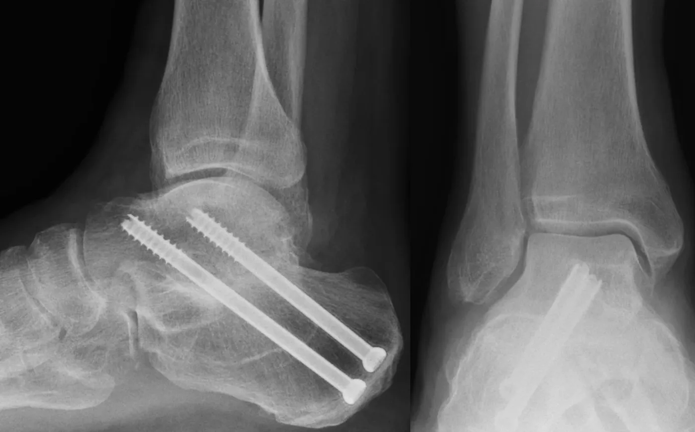
Hindfoot fusion treats severe arthritis, deformity or instability at the back of the foot, most commonly the subtalar, talonavicular and/or calcaneocuboid joints. It may be a subtalar, double, triple or TTC (subtalar and ankle) fusion. Damaged cartilage is removed, alignment corrected and the bones fixed with screws, staples, nails or plates while they fuse. You stay in hospital briefly, are non-weight-bearing in a cast or boot at first, and X-rays at 6 to 12 weeks guide the return to weight-bearing.
- Anaesthetic
- General or regional anaesthetic
- Hospital stay
- Short inpatient stay
- Walking afterwards
- Non-weight-bearing with crutches at first, in a cast or boot
- Full recovery
- 3 to 6 months and beyond, depending on joints fused
Why is surgery sometimes needed?
Hindfoot fusion surgery is used to treat severe arthritis, deformity or instability affecting the back of the foot, most commonly involving the subtalar, talonavicular and/or calcaneocuboid joints. These conditions can cause persistent pain, swelling and difficulty walking, particularly on uneven ground. Fusion stabilises the affected joints, removes painful movement and helps restore a more comfortable and stable foot.
What does the operation involve?
Hindfoot fusion may involve one or more joints depending on your condition. Common procedures include:
- Subtalar fusion
- Double fusion (two joints)
- Triple fusion (subtalar, talonavicular and calcaneocuboid joints)
- TTC fusion (subtalar and ankle joints)
The operation typically involves:
- A short inpatient stay, under general or regional anaesthetic
- Removal of damaged cartilage from the affected joints
- Correction of foot alignment
- Fixation with metalwork (screws, staples, nails or plates) to hold the bones while they fuse
- Gradual bone healing over several months to form a solid, pain-free unit
Prof Mason will discuss which joints need fusion and why.
Recovery: what to expect
0 to 2 weeks
- Your foot will be placed in a cast or rigid boot
- Non-weight-bearing with crutches is common initially
- Elevation is important to reduce swelling
- Stitches removed around 10 to 14 days
2 to 6 weeks
- Continued immobilisation in a cast or boot
- Gentle movement of non-fused joints may begin
6 to 12 weeks
- X-rays are taken to assess fusion progress
- Gradual progression to partial weight-bearing in a boot if healing allows
- Physiotherapy may start to maintain strength and improve gait
3 to 6 months and beyond
- Progression to full weight-bearing as the fusion consolidates
- Transition into supportive footwear
- Ongoing improvement in comfort and walking ability; swelling gradually reduces
- Return to low-impact activities such as walking, cycling or golf
Recovery varies with the exact procedure and with each person. You will get a plan specific to your operation.
Expected outcomes
Before surgery
- Persistent hindfoot pain
- Difficulty walking on uneven ground
- Swelling and instability
- Reduced activity and confidence
After surgery
- Significant reduction in pain
- Improved stability and alignment
- More comfortable walking
- Improved confidence in daily activities
- Long-lasting results once fusion is complete
Most patients achieve solid fusion and durable pain relief. Some loss of hindfoot motion is expected but usually well tolerated. Supportive footwear helps protect adjacent joints long term. Recovery time depends on the number of joints fused and your individual healing response.
Consent information
Read the consent form before your operation so you know what you are agreeing to. Bring any questions to your pre-operative appointment.
- Consent form: subtalar fusion (Word document)
- Consent form: double fusion (Word document)
- Consent form: triple fusion (Word document)
- Consent form: TTC (tibiotalocalcaneal) fusion (Word document)
Considering this operation?
Talk it through with Prof Mason. New consultation £250.

Consultant Foot and Ankle Surgeon, MB BCh, PhD, FRCS (Tr&Orth), FRCS (Glasg), SFHEA, FFSTEd. GMC 6074953.
Prof Mason's published research on this topic
Peer-reviewed papers by Prof Mason and his colleagues. Links open the paper or its PubMed record.
- Widnall J, Mason LW, Molloy A. Medial approach to the subtalar joint. Foot and Ankle Clinics, 2018.
- Durston A, Baboo R, Kadambande S, Hariharan K, Mason LW. Minimally invasive calcaneal osteotomies: are neurovascular structures at risk? A cadaveric study. Journal of Foot and Ankle Surgery, 2015.