In short
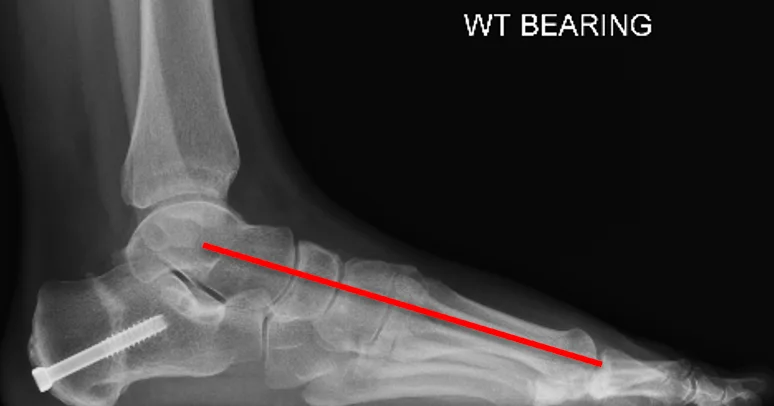
Flat foot reconstruction treats a painful or progressive flat foot that has not improved with insoles and physiotherapy. It is usually a combination of procedures chosen for your foot: repairing or transferring the tibialis posterior tendon, realigning bones such as the heel (osteotomy), fusing unstable or arthritic joints and rebalancing the ligaments. You keep weight off the foot in a cast or boot at first, start protected walking from about 6 weeks, and function supports daily activities after about 6 months.
- Anaesthetic
- General or regional anaesthetic
- Walking afterwards
- No weight on the foot at first, in a cast or boot
- Full recovery
- 6 to 12 months
What is flat foot reconstruction?
It is surgery for a painful or progressive flat foot (often called adult-acquired flat foot or progressive collapsing foot deformity) that has not improved with non-surgical care. The aims are to restore the arch, improve alignment and relieve pain from tendon failure, arthritis or structural collapse.
What does the operation involve?
Usually a combination of procedures, planned for your individual foot. Prof Mason will go through the exact plan with you.
- Tendon procedures: repairing the tibialis posterior tendon, or transferring a neighbouring tendon to take over its job
- Osteotomies: cutting and realigning bones, such as moving the heel bone inwards or adjusting the midfoot
- Fusion (arthrodesis): joining unstable or arthritic joints
- Ligament procedures: reconstructing the spring ligament and balancing the soft tissues
- Calf lengthening if a tight calf is contributing
The recovery depends on which procedures are combined, how many joints are involved and your fitness beforehand.
Recovery: what to expect
0 to 2 weeks
- A cast or boot protects the reconstruction
- No weight on the foot, using crutches
- Keep the foot raised to reduce swelling
- Stitches are usually removed at 10 to 14 days
2 to 6 weeks
- Continued protection in a boot or cast
- X-rays may be taken to check early healing
- Gentle non-weight-bearing movement and strengthening exercises may begin
6 to 12 weeks
- Protected weight-bearing starts, partial then full, in a boot or supportive shoe as advised
- Physiotherapy works on walking pattern, movement and strength
3 to 6 months and beyond
- Move into normal footwear with arch support
- Walking distance and daily activities increase gradually
- Some swelling may persist but keeps reducing
- By about 6 months, function usually supports daily activities; low-impact sport is often possible
Recovery varies with the exact procedure and with each person. You will get a plan specific to your operation.
Expected outcomes
Before surgery
- Pain in the arch and inner ankle with walking or standing
- Difficulty wearing normal shoes
- Swelling or stiffness across the arch
- Reduced activity because of discomfort
After surgery
- Significant reduction or resolution of pain
- Better stability and alignment of the foot
- More comfortable walking
- Supportive footwear can be worn comfortably
Most patients achieve a stable correction and lasting pain relief. Nearby joints usually adapt well, though a small percentage may experience increased stress on them in the long term. Supportive shoes and a gradual increase in activity help the result.
Consent information
Read the consent form before your operation so you know what you are agreeing to. Bring any questions to your pre-operative appointment.
- Consent form: flat foot reconstruction (Word document)
- Consent form: hindfoot osteotomy (heel bone realignment) (Word document)
- Consent form: pes cavus (high arch) reconstruction (Word document)
Considering this operation?
Talk it through with Prof Mason. New consultation £250.

Consultant Foot and Ankle Surgeon, MB BCh, PhD, FRCS (Tr&Orth), FRCS (Glasg), SFHEA, FFSTEd. GMC 6074953.
Prof Mason's published research on this topic
Peer-reviewed papers by Prof Mason and his colleagues. Links open the paper or its PubMed record.
- Heyes G, Swanton E, Vosoughi AR, Mason LW, Molloy AP. Comparative study of spring ligament reconstructions using either hamstring allograft or synthetic ligament augmentation. Foot & Ankle International, 2020.
- Swanton E, Fisher L, Fisher A, Molloy A, Mason LW. An anatomic study of the naviculocuneiform ligament and its possible role maintaining the medial longitudinal arch. Foot & Ankle International, 2019.
- Chapman J, Dalal N, Molloy A, Jenkinson H, Singh A, Sirikonda S, Mason L. Association of fusion of the first metatarsophalangeal joint and pes planus deformity correction. Foot & Ankle International, 2023.
- Heyes G, Vosoughi AR, Weigelt L, Mason LW, Molloy AP. Pes planus deformity and its association with hallux valgus recurrence following Scarf osteotomy. Foot & Ankle International, 2020.